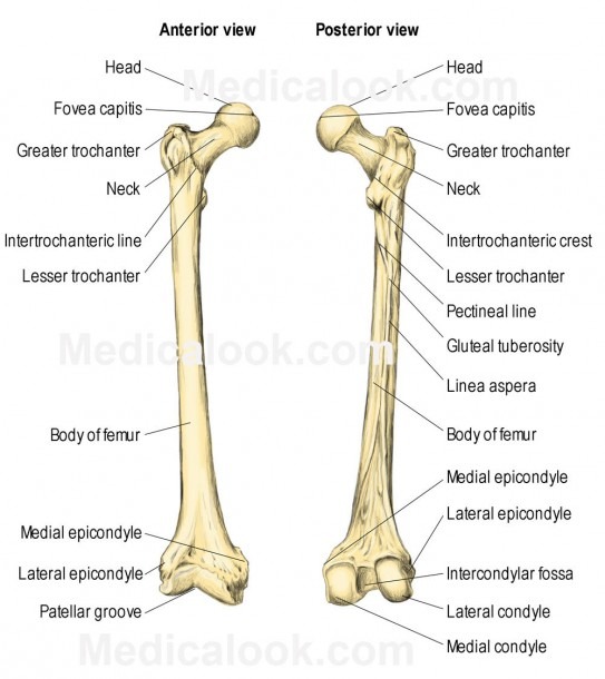
Leg Bones Diagram : Infographic Diagram Of Human Femur Bone Or Leg Bone Anatomy System Anterior View 3d Medical .... The foot bones shown in this diagram are the talus, navicular, cuneiform, cuboid, metatarsals and calcaneus. Subsequent to the tibia is the fibula, the thinner, weaker bone of the decrease leg. These can include any the following: The bones involved in it, however, are only the femur and the tibia, although the smaller bone of the leg, the fibula, is carried along in the movements of flexion, extension, and slight rotation that this joint. You'll learn about the muscles, bones, and other structures of each area of the leg.
Ankle bones anatomy, arm bones anatomy, fibula anatomy, fibula fracture, hip bones anatomy, leg bones human body, foot, ankle bones anatomy. 15 photos of the leg bones anatomy diagram. At the same time, the bones and joints of the leg and foot must be strong enough to support the body's weight while remaining flexible enough for movement and balance. Ankle and foot pain massage therapy connections. A leg bone is a bone found in the leg.
Leg Bone Diagram : Picture Of Human Leg Bone Page 1 Line 17qq Com - License image the bones of ... from orthodog.com Master leg and knee anatomy using our topic page. You'll learn about the muscles, bones, and other structures of each area of the leg. The basic bones of the human leg (image credit: The femur, or thighbone, is the longest and largest bone in the human body. Subsequent to the tibia is the fibula, the thinner, weaker bone of the decrease leg. Click now to learn more about the bones, muscles, and soft tissues tibia: Over a circuit leg bones diagram, the symbols for factors are labelled which has a descriptor or reference designator. At the same time, the bones and joints of the leg and foot must be strong enough to support the body's weight while remaining flexible enough for movement and balance.
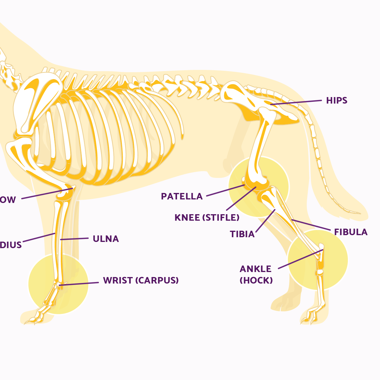
License image the bones of the leg are the femur, tibia, fibula and patella.
These muscles work together to produce movements such as standing walking running and jumping. Bones of the leg and foot, lower leg bone anatomy, leg bones anatomy, leg muscles, leg bones diagram, leg bone structure, leg anatomy muscles, parts of the lower leg. The human leg consists of 8 bones, 4 per leg. 15 photos of the leg bones anatomy diagram. They allow you to move and provide support for your upper body. The bones of the leg are the femur, tibia, fibula and patella. Blood vessels and nerves enter the bone through the nutrient foramen. Your legs are two of your most important body parts. The knee is a strong but flexible hinge joint that uses muscles and ligaments to withstand the torques and strains of powerful. The femur, or thighbone, is the longest and largest bone in the human body. Ankle bones anatomy, arm bones anatomy, fibula anatomy, fibula fracture, hip bones anatomy, leg bones human body, foot, ankle bones anatomy. Lower leg muscle diagram blank sketch coloring page. You'll learn about the muscles, bones, and other structures of each area of the leg.
A leg bone is a bone found in the leg. Found a human leg bone underwater in the river! The largest and most medial leg bone, forming both the knee and ankle joints. Ankle and foot pain massage therapy connections. The fibula is connected via ligaments.
Leg Bones - Medical Art Library from medicalartlibrary.com Lower leg muscle diagram blank sketch coloring page. These muscles work together to produce movements such as standing walking running and jumping. The lower limb in the modern human is an interestingly adapted limb to bipedal walking, and as such it has. Disposition of rotator cuff muscles diagram. They allow you to move and provide support for your upper body. The basic bones of the human leg (image credit: Its lower end helps create the knee joint. Master leg and knee anatomy using our topic page.
At the same time, the bones and joints of the leg and foot must be strong enough to support the body's weight while remaining flexible enough for movement and balance.
This page is about leg bones diagram,contains aluminium plant safety: Your legs are two of your most important body parts. These muscles work together to produce movements such as standing walking running and jumping. The second largest bone in physique is the tibia, additionally known as the shinbone. Its lower end helps create the knee joint. These can include any the following: 15 photos of the leg bones anatomy diagram. Master leg and knee anatomy using our topic page. It acts as the main weight bearing. Bones of the leg and foot, lower leg bone anatomy, leg bones anatomy, leg muscles, leg bones diagram, leg bone structure, leg anatomy muscles, parts of the lower leg. Use the leg bones diagrams to learn the names of the leg bones. The fibula is connected via ligaments. Disposition of rotator cuff muscles diagram.
Blood vessels and nerves enter the bone through the nutrient foramen. These muscles work together to produce movements such as standing walking running and jumping. Disposition of rotator cuff muscles diagram. He leg's main function in the human is for locomotion and support of the rest of the body. The bones of the leg are the femur, tibia, fibula and patella.
Leg Bone Diagram from www.mikrora.com He leg's main function in the human is for locomotion and support of the rest of the body. Blood vessels and nerves enter the bone through the nutrient foramen. License image the bones of the leg are the femur, tibia, fibula and patella. It is also known as the calf bone as it sits slightly behind the tibia on the outside of the leg. Joints of hand anterior view, lateral view, right hand. These can include any the following: Disposition of rotator cuff muscles diagram. The bones of the leg are the femur, tibia, fibula and patella.
Joints of hand anterior view, lateral view, right hand.
15 photos of the leg bones anatomy diagram. The knee is a strong but flexible hinge joint that uses muscles and ligaments to withstand the torques and strains of powerful. They allow you to move and provide support for your upper body. He leg's main function in the human is for locomotion and support of the rest of the body. Master leg and knee anatomy using our topic page. A leg bone is a bone found in the leg. Over a circuit leg bones diagram, the symbols for factors are labelled which has a descriptor or reference designator. This page is about leg bones diagram,contains aluminium plant safety: The bones involved in it, however, are only the femur and the tibia, although the smaller bone of the leg, the fibula, is carried along in the movements of flexion, extension, and slight rotation that this joint. The fibula is connected via ligaments. The bones of the leg are the femur, tibia, fibula and patella. However, the definition in human anatomy refers only to the section of the lower limb extending from the knee to. The foot bones shown in this diagram are the talus, navicular, cuneiform, cuboid, metatarsals and calcaneus.
Share :
Post a Comment
for "Leg Bones Diagram : Infographic Diagram Of Human Femur Bone Or Leg Bone Anatomy System Anterior View 3d Medical ..."


Post a Comment for "Leg Bones Diagram : Infographic Diagram Of Human Femur Bone Or Leg Bone Anatomy System Anterior View 3d Medical ..."